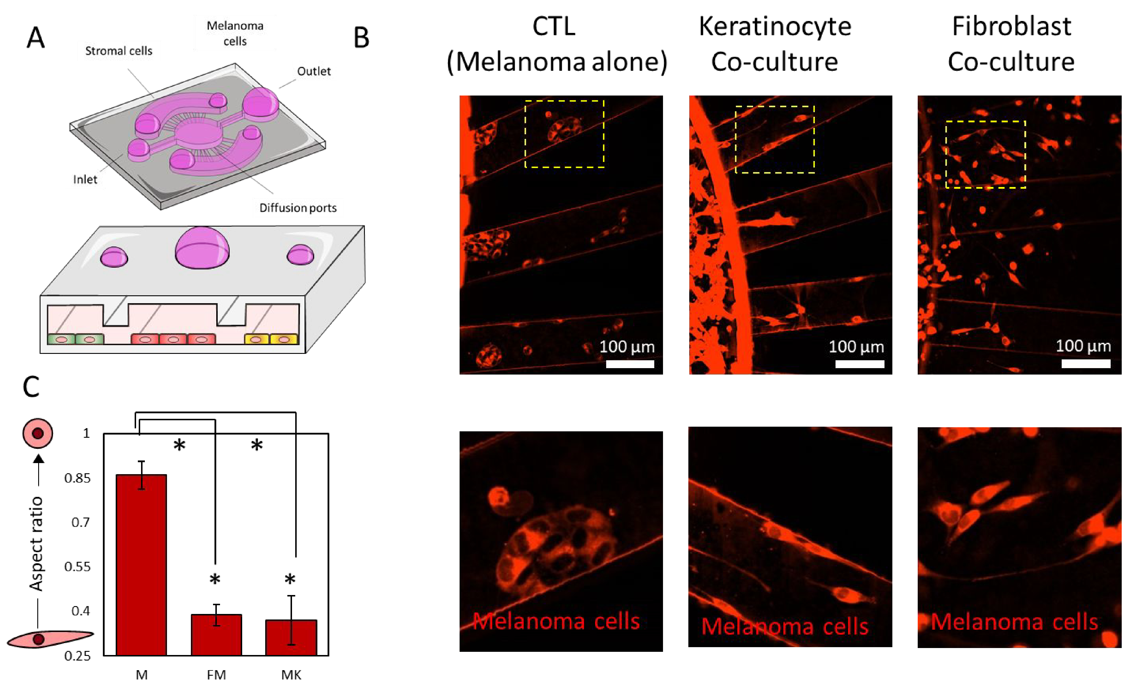
Figure 2. Morphological analysis.

A) Scheme illustrating the experimental protocol: melanoma cells were confined in the central chamber using the air-wall method with/without dermal fibroblasts/keratinocytes in the lateral chambers. After 3 days in culture, the microdevices were imaged to visualize cell migration and morphology. B) Fluorescence microscopy images showed melanoma cells stained in red after 3 days in culture in monoculture (control condition) and in the presence of fibroblasts, or keratinocytes, or both in the lateral chambers. When cultured alone, melanoma cells formed multicellular clusters. When cultured in the presence of fibroblasts, keratinocytes, or both, melanoma cells spread and appeared as isolated cells. C) Graph showed the melanoma cell aspect ratio in monoculture and in the presence of dermal fibroblasts and keratinocytes. Asterisks denote p-value<0.05. Experiments were repeated at least three independent times with replicates showing showed similar trends.
