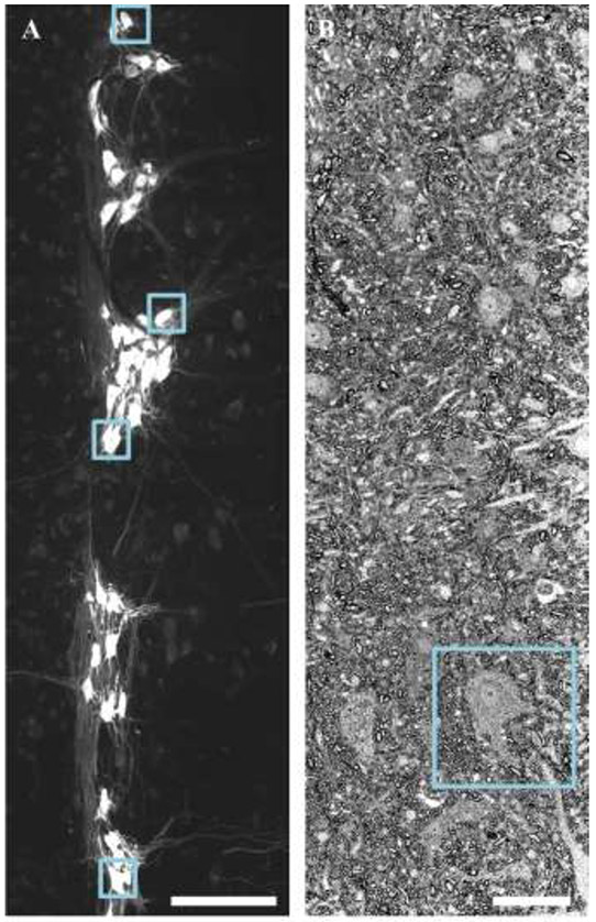
Figure 1: Identification and sampling of motor neurons.
A shows specific labeling of phrenic motor neurons via retrograde labeling with Alexa 488-conjugated CTB is readily apparent in z-projection images of horizontal sections (100 μm thick) of cervical (~C4-C5 pictured) spinal cord. The blue squares show every 20th phrenic motor neuron, which was then assessed for mitochondrial volume density. B shows a survey section (~10 μm thick) containing a putative phrenic motor neuron (inside blue square). These putative phrenic motor neurons later underwent serial block face EM. Scale Bars: A=200 μm; B=20 μm.

