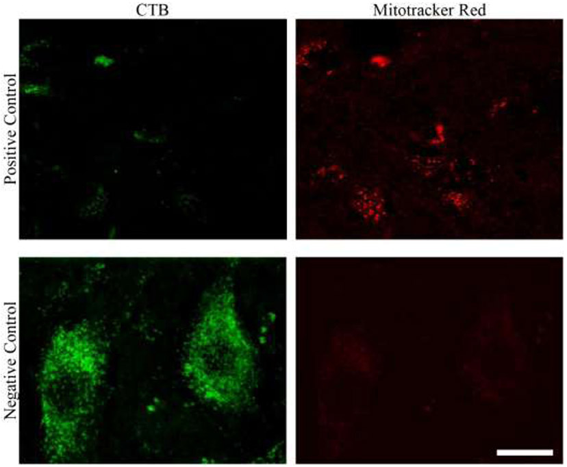
Figure 12: Positive and negative controls.
The top row shows a portion with an absence of labeled phrenic motor neurons (left), albeit with a reliable MitoTracker Red fluorescent signal. The bottom row shows little non-specific fluorescence on the 575-630 nm channel when an Alexa 488-conjugated labeled phrenic motor neuron was imaged without any intrathecal application of MitoTraker Red (i.e., a negative control). Scale Bar: 5 μm.

