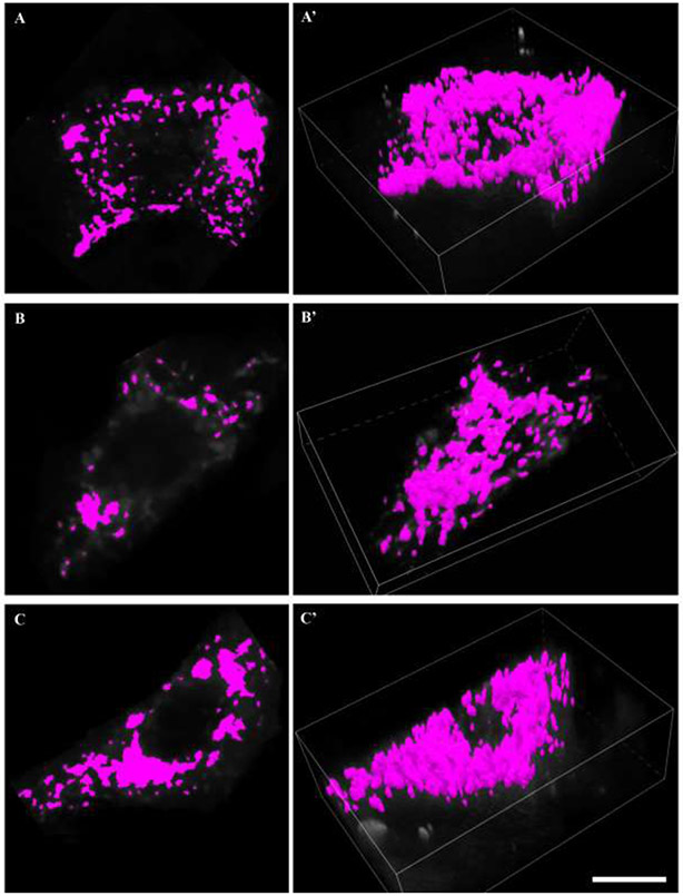
Figure 5: Mitochondrial volume reconstructions.
A, B and C show binarisation of deconvolved MitoTracker red signal within a mid-nuclear section of a labelled phrenic motor neuron soma. A’, B’ and C’ show respective mitochondrial volume reconstructions in 3D of the all mitochondrial elements within the labeled phrenic motor neuron. Scale Bar: 10 μm.

