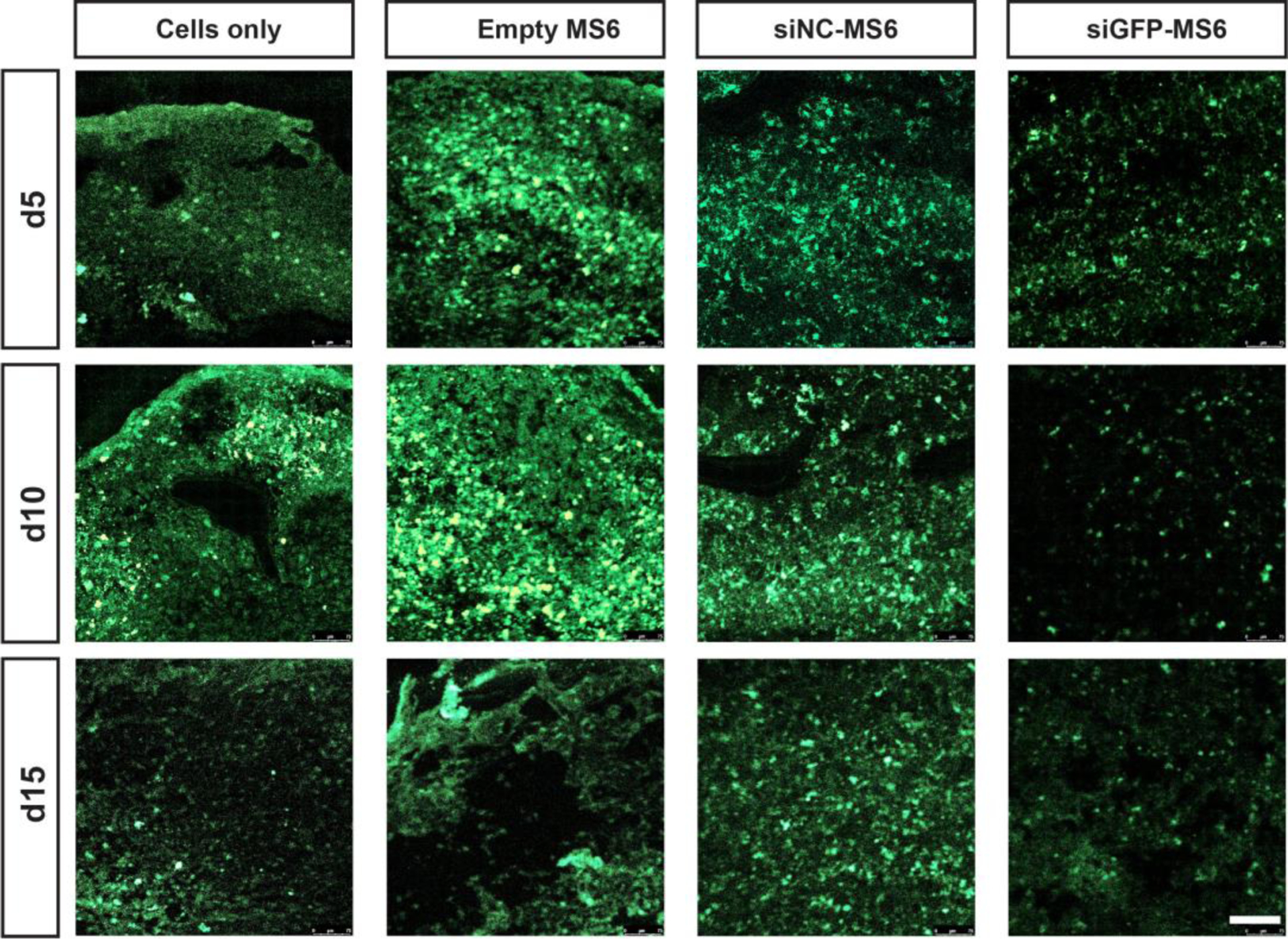
Figure 8.

siGFP-mediated silencing of GFP expression within GFP-hMSC aggregates. Tissue sections of aggregates show GFP signal (green) of GFP-hMSC aggregates incorporated with cells only, and empty-, siNC- and siGFP-MS6 at different culture times. Scale bar indicates 100 μm.
