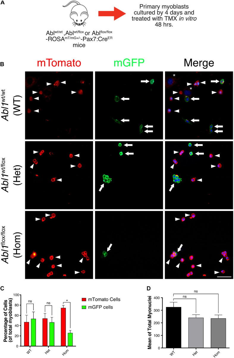
FIGURE 7.
GFP positive primary myoblasts population decrease in c-Abl Knockout cells recombined in vitro. (A) Protocol to induce Abl1 recombination in vitro. Primary myoblasts were isolated from Abl1WT/WT (WT), Abl1wt/flox (Het), or Abl1flox/flox (Hom) ROSAmT/mG-Pax7creERT2 mice and maintained 4 days in PM. During this time, cells were treated with vehicle or Tamoxifen (TMX, 10 μM) for 48 h. After that, cells were fixed, and direct fluorescence was detected. Nuclei were stained with Hoechst 33342 (blue). (B) Arrowheads show mTomato positive cells and arrows show mGFP positive cells. The asterisk shows non-myogenic cells. Scale bar: 50 μm. (C) Quantification of the percentage of mTomato positive and mGFP positive myoblasts from (B) (n = 3, *P-value < 0.05, multiple t-Student test, ns, not significant). (D) Total number of myoblasts do not change in different mice. Mean of total myoblasts was determined for every mouse from (B). WT mean: 324.5 ± 39.50; Het mean: 239.7 ± 25.06; Hom mean: 234.7 ± 28.30 (n = 3, P-value > 0.05, ANOVA test, ns, not significant).

