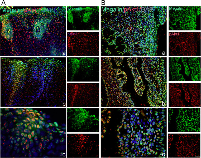
Fig. 5.
Megalin-positive cells express phosphorylated Akt-1/protein kinase B. Cells expressing megalin (green staining) and phospho-AKT1/protein kinase B (pAkt1) (red staining) were detected by the use of anti-megalin and anti-pAkt1 (phospho-Thr308) antibodies in paraffin-embedded sections of the cervical tissue samples, classified as HSIL (CIN2) (A) or CIN3/CIS (B). Blue marks DAPI staining of nuclei and yellow marks the overlapping of megalin with pAkt1. Scale bars: 50 μm (A a, b; B a–c) and 20 μm (A c)

