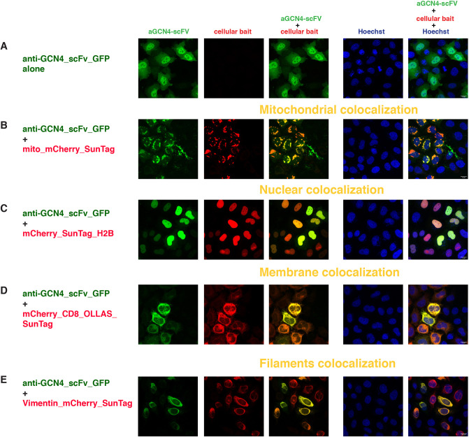
Fig. 2.
Intracellular binding of anti-GCN4 scFv (SunTag system). Confocal images of HeLa cells transiently transfected with (A) anti-GCN4_scFv_GFP alone or with (B-E) the combination of anti-GCN4_scFv_GFP and (B) mito_mCherry_SunTag, (C) mCherry_SunTag_H2B, (D) mCherryCD8_OLLAS_SunTag or (E) vimentin_mCherry_SunTag. The first column represents the GFP channel (green), the second column is the mCherry channel (red), the third column is the overlay of the two channels, showing the colocalization (indicated in yellow) of the anti-GCN4_scFv with the respective mitochondrial (B), nuclear (C), membrane (D) and filament (E) baits; the fourth column represents the nuclear Hoechst staining (blue) and the fifth column is the merge of all three channels. Scale bars: 15 µm. Images were taken 24 h post-transfection. Transfected constructs are indicated at the left of each row and the single and merge channels are indicated at the top of the respective columns. The figures are from a representative experiment, performed at least three times.

