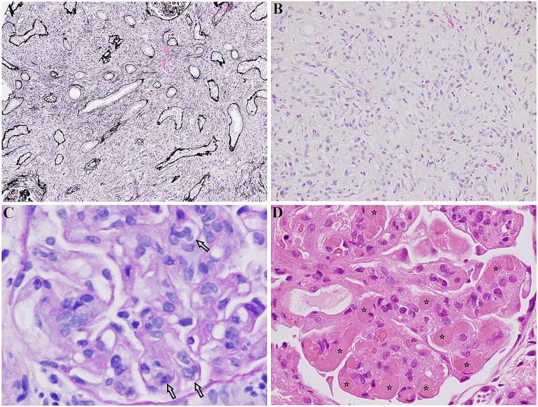
Fig. 5.
a IgG4-related tubulointerstitial nephritis: tubules separated by expansile interstitial fibrosis and inflammation. Jones methenamine silver, × 4. This image was originally used in Kidder et al. [22]. b IgG4-related tubulointerstitial nephritis: interstitial “storiform” fibrosis and inflammation. Haematoxylin and eosin stain, × 40. This image was originally used in Kidder et al. [22]. c Membranoproliferative glomerulonephritis in Sjögren’s: glomerulus showing intracapillary hypercellularity (indicated by arrows) (PAS, × 400). This image was originally used in Kidder et al. [22]. d Cryoglobulinaemic glomerulonephritis in Sjögren’s: glomerulus containing several hyaline thrombi, “cryoplugs” (indicated by stars), in capillaries. H + E, × 400. This image was originally used in Kidder et al. [22].

