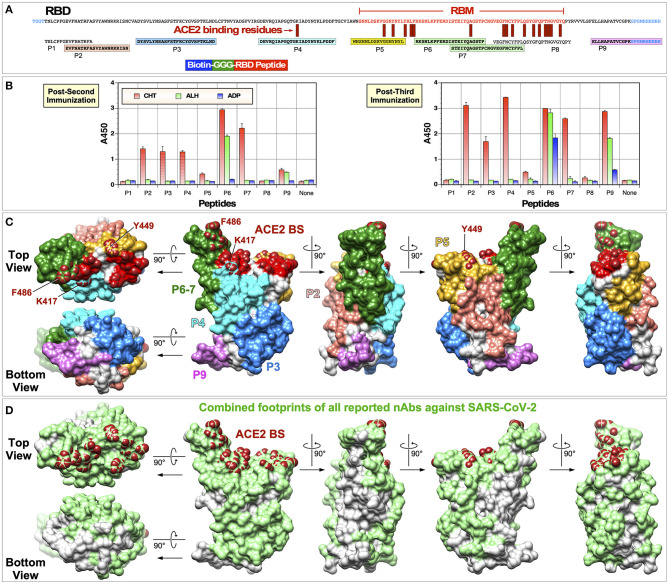
Figure 8.
Identification of immunogenic epitopes using biotinylated peptides. (A) Sequences of the RBD and biotinylated peptides used are shown. Amino acid residues located within the RBM are indicated in red text. Immunogenic epitopes are highlighted in colored boxes. (B) ELISA was done using serum samples collected 2 weeks after the second or third immunization (1:300 dilution). ELISA plates were coated with streptavidin (300 ng per well) and 100 ng peptides were attached to streptavidin. Negative controls without any peptides are indicated as “None.” (C) Locations of immunogenic peptides are mapped onto the RBD structure (PDB: 6M0J). (D) Combined footprints of 32 neutralizing mAbs with known crystal structures are shown.

