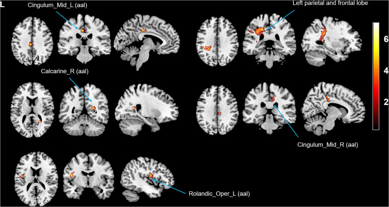
FIGURE 3.
Intergroup differences in WMV between the tinnitus patients before and after sound therapy. Compared with baseline, after sound therapy, the patients with tinnitus exhibited increased WMV in the cingulum (cingulate), right calcarine, left rolandic operculum, and left parietal and frontal lobes (corrected at the non-stationary cluster level with FWE p < 0.05). FWE, family wise error, WMV, white matter volume. The color bar represents the extent of reduction in WMV.

