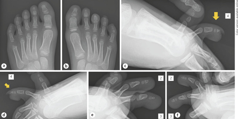
Fig. 2.
Plain radiography at the time of the initial assessment. The distal phalanges of the fourth toes were slightly small and dorsally deviated. a Dorsal view of the left foot. b Dorsal view of the right foot. c Lateral view of the left fourth toe (arrow: fourth toe). d Lateral view of the right fourth toe (arrow: fourth toe). e Lateral view of the left second and third toes. f Lateral view of the right second and third toes.

