Figure 3.
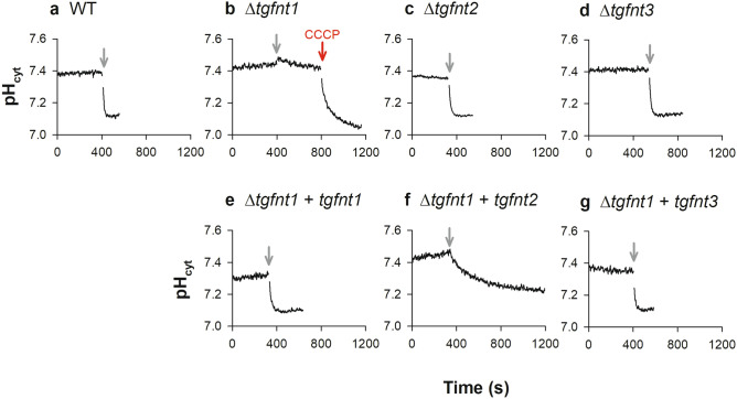
Measurements of the effect of l-lactate on cytosolic pH (pHcyt) reveal a role for TgFNT1 in lactate transport across the parasite plasma membrane. Extracellular tachyzoites were loaded with BCECF and suspended in Saline Solution at 4 °C. The grey arrows denote the addition of 10 mM l-lactate. The red arrow denotes the addition of the protonophore CCCP (10 µM). The top panels show data for Δtgfnt1 (b), Δtgfnt2 (c), Δtgfnt3 (d) parasites and their wild-type parental parasites (a). The bottom panels show data for Δtgfnt1 parasites complemented with tgfnt1 (e), tgfnt2 (f) or tgfnt3 (g). The traces are from a single experiment for each parasite line, and are representative of those obtained in at least three independent experiments. The time taken to reach a minimum pHcyt value after the addition of l-lactate was > 400 s in all experiments for Δtgfnt1 parasites complemented with tgfnt2, compared to < 150 s in all experiments for WT parasites, Δtgfnt2 parasites, Δtgfnt3 parasites, and Δtgfnt1 parasites complemented with tgfnt1 or tgfnt3.

