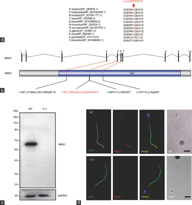
Figure 2.
The impact of the MNS1 variant. (a) Evolutionary conservation analysis of the MNS1 variant among different species. (b) The positions of the identified variant (red) and previously reported mutations (black) in MNS1 in patients are shown. The trichohyalin–plectin–homology (TPH) domain (http://smart.embl-heidelberg.de/) in MNS1 is indicated in blue. (c) Western blot analysis shows that the MNS1 protein (approximately 60 kDa) is absent from the proband's sperm. (d) Immunostaining analysis shows that MNS1 is localized in the flagella of the sperm in the NC, but was absent in the sperm of the proband (IV-1; n = 120). In the NC, the staining of MNS1 in the midpiece is prominent, whereas that in the principal and end piece is distributed discretely with distinct dots. Scale bars = 5 μm. MNS1: meiosis-specific nuclear structural 1; α-tub: alpha-tubulin; GAPDH: glyceraldehyde 3-phosphate dehydrogenase; NC: normal control.

