Figure 3.

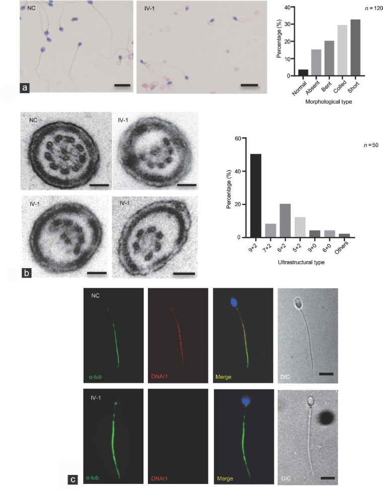
The morphological and structural abnormalities in the proband with MNS1 mutation. (a) H&E staining shows that there were many morphological abnormalities in the proband's sperm, including absent, bent, coiled, and short flagella, compared to the NC. Scale bars = 10 μm. A histogram summary of the sperm morphology is shown on the right. (b) Transmission electron micrographs show subtle ultrastructural defects in the proband's sperm, with the absence of outer doublet microtubules or central pair complex of the cross-sections, compared to NC samples. A red asterisk shows the absence of central pair complex. Scale bars = 100 nm. A histogram summary of the ultrastructural defects is shown in the right. (c) Immunofluorescence shows the expression and location of DNAI1 (a marker for outer dynein arm). Compared to NC, DNAI1 staining appears absent from the sperm flagella in the proband (n = 120). Scale bars = 5 μm. MNS1: meiosis-specific nuclear structural 1; α-tub: alpha-tubulin; GAPDH: glyceraldehyde 3-phosphate dehydrogenase; NC: normal control; H&E: hematoxylin and eosin.
