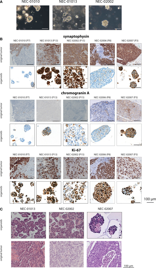
Figure 1.
Histomorphology of GEP-NEC organoid lines and matching original tumor. (A) Phase-contrast photomicrographs of GEP-NEC organoid lines. Scale bar = 100 µm. (B) Immunostainings of synaptophysin, chromogranin and Ki-67 for organoids and original tumor. P7 indicates passage 7. (C) Hematoxylin and eosin (H&E) stained slides of GEP-NEC organoid lines and original tumors. NEC-01013: passage (P) 12; NEC-02002: P20; NEC-02007: P3.

