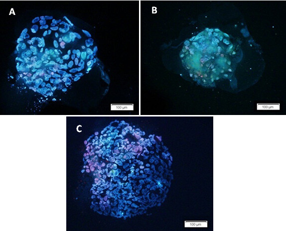
Figure 2.

Hoechst and PI staining of the blastocysts as a valuable approach for assessment of the number of viable and dead cells. The figure shows three blastocysts developed from different sources: A) from twin A embryo after in vitro twinning; B) from twin B embryo after in vitro twinning; C) from embryo without any manipulation. The nuclei of viable cells appear blue, while dead cells are seen as red.
