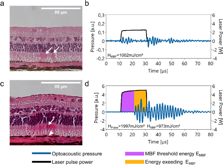
Figure 7.
(a) HE-stained image of an ophthalmoscopically nonvisible region. A slightly stronger eosin staining in the photoreceptor inner segments is noticed (arrows). A maximal temperature of 79.5 ± 3.9°C was determined for this lesion from the transient. (b) Laser pulse and optoacoustic transient acquired during the irradiation process of the region displayed in (a); MBF was not detected. (c) HE-stained image of an ophthalmoscopically faintly visible region. Remarkable changes are an eosinophilic unstructured substance directly on the RPE (arrowhead), a stronger staining of eosin in the photoreceptor inner segments (arrow), and the deformation of a photoreceptor cell layer (asterisk). (d) Laser pulse and optoacoustic transient acquired during the irradiation process of the region displayed in subfigure c, revealing MBF.

