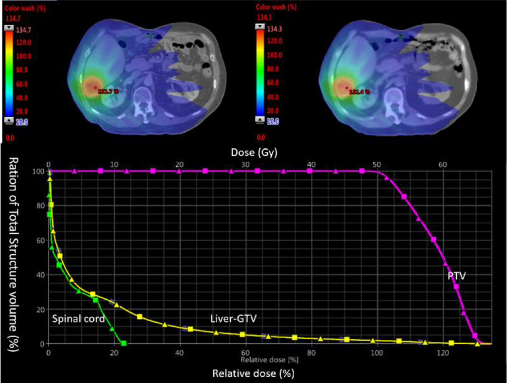
Figure 10.

Top: dose distribution on CT (left) and synthetic CT (right) images. Bottom: dose volume histograms of PTV, liver (exclude gross tumor volume) and spinal cord from CT (squares) and synthetic CT (triangles) calculations.

Top: dose distribution on CT (left) and synthetic CT (right) images. Bottom: dose volume histograms of PTV, liver (exclude gross tumor volume) and spinal cord from CT (squares) and synthetic CT (triangles) calculations.