Figure 6.

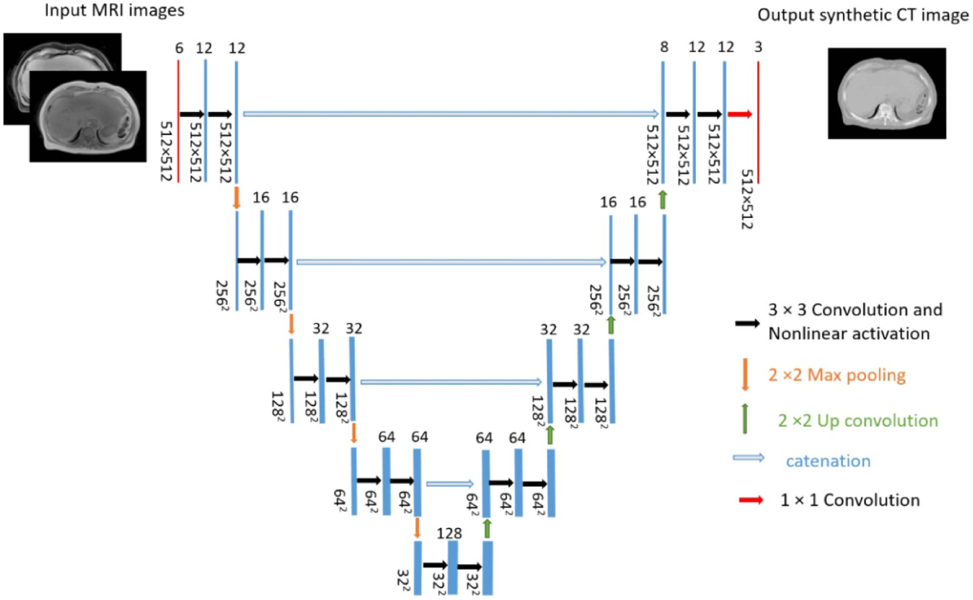
U-net architecture. For simplicity, the central of three contiguous input (T1-weighted in-phase and water images from Dixon MRI) and output (‘semi-synthetic’ CT image) channels are displayed.

U-net architecture. For simplicity, the central of three contiguous input (T1-weighted in-phase and water images from Dixon MRI) and output (‘semi-synthetic’ CT image) channels are displayed.