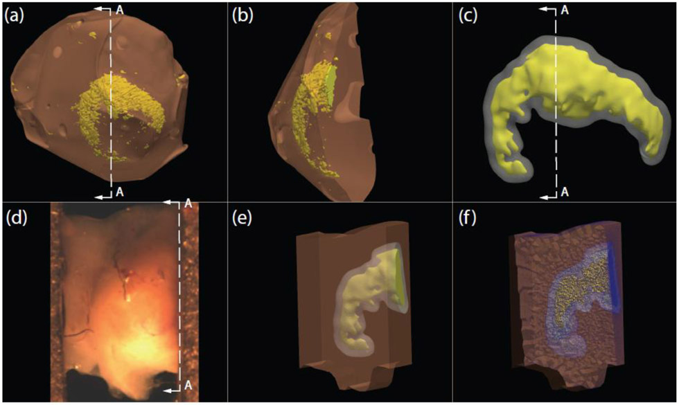
Fig. 1.
(a) Surface meshes of cerebral aneurysm dome tissue and calcification created from 3D micro-CT protocol. (b) Cross-section A-A of aneurysm dome and calcification surface meshes (c) Smoothed calcification surface mesh in yellow, and the offset surface that models the boundary of near-calcification region (NCR) in grey. (d) Experimental uniaxial tissue specimen at zero load configuration. (e) Opaque specimen solid model illustrating internal calcification in yellow and near-calcification region surrounding the calcification. (f) Cross-section image of the 3D finite element mesh of the in-situ uniaxial specimen

