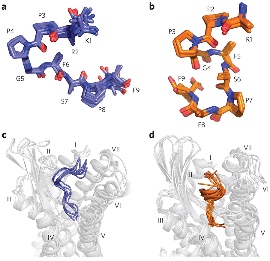
Figure 3 ∣. Backbone structures of DAKD in complex with human B1R in comparison to BK bound to human B2R.
Only backbone and Cβ atoms are shown. (a) The backbone structure of DAKD calculated from NMR data features a V-shaped fold with a β-turn-like structure around P3–F6. (b) The NMR-based backbone structure of BK is characterized by an overall S-shape with a 310-helix-like segment (P2–F5) in the middle. (c,d) Rosetta modeling of DAKD in B1R (c) and BK in B2R (d) reproduces the characteristic V-shape of DAKD and the S-shape fold of BK (see text and Supplementary Fig. 10 for further details).

