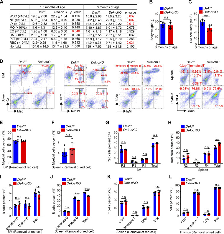
Figure S1.
DEK deletion impairs hematopoiesis in mice. (A) PB complete blood cell counts of Dekfl/fl and Dek-cKO mice at 1.5 and 3 mo of age (n = 5–7). WBC, white blood cells; NE, neutrophils; LY, lymphocytes; MO, monocytes; EO, eosinophils; BA, basophils; PLT, platelets; Hb, hemoglobin. (B and C) Weight analysis and BM cells count in Dekfl/fl and Dek-cKO mice at 3 mo of age (n = 6). (D) FACS analysis of myeloid cells (Mac+Gr1+), red cells (R1: Ter119medCD71high; R2: Ter119highCD71high; R3: Ter119highCD71med; R4: Ter119highCD71low), B cells (immature B: IgM−B220+; mature B: IgM+B220+), and T cells (immature T: CD8a+CD4+; CD4+ T; CD8+ T) in BM, spleen, and thymus of Dekfl/fl and Dek-cKO mice at 3 mo of age. (E and F) Percent analysis of myeloid cells in BM and spleen cells (n = 5). (G and H) Percent analysis of red cells in BM and spleen cells (n = 5). (I and J) Percent analysis of B cells in BM and spleen cells (n = 5). (K and L) Percent analysis of T cells in spleen and thymus cells (n = 5). Error bars represent means ± SD. **, P < 0.01; ***, P < 0.001; Student’s t test. Data are representative of three independent experiments.

