
Fig. 4.
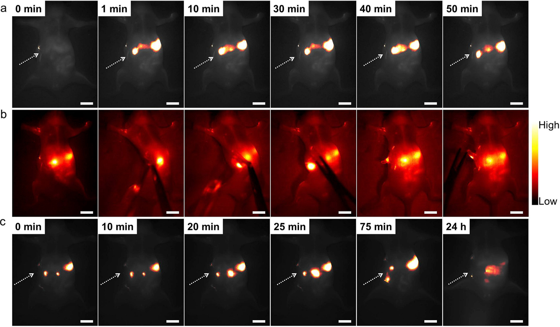
(a) Representative in vivo NIR-II fluorescence images of a BALB/c nude mouse with intestinal obstruction. Compared with GI tract images, DCNP@PDA NPs still accumulated at the obstructive sites even at 50 min post-gavage. (b) In vivo NIR-II FI-guided intestinal obstruction mapping on BALB/c nude mice. (c) In vivo NIR-II FI of GI tract after intestinal obstruction surgery. DCNP@PDA NPs passed through the obstructive site and discharged successfully after 24 h post-gavage. Scale bars are 10 mm.