
Figure 1.
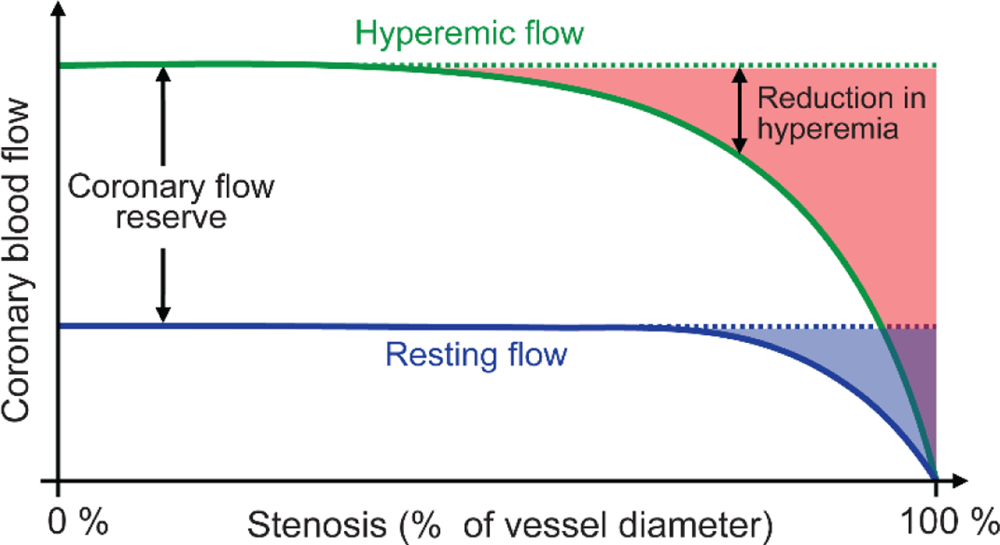
Relationship between percent narrowing of the coronary artery diameter (stenosis, x-axis) and coronary blood flow (y-axis) during rest (dashed blue line) and hyperemia (solid green line). The resting flow is maintained at normal levels (dotted black line) up to 85–90% stenosis because of autoregulation. During hyperemia, however, increases in flow are attenuated at 50% or greater stenosis, with more severe stenosis resulting in greater attenuation of hyperemia flow. The shaded blue region denotes the decrease in resting flow d the shaded red region denotes reduction in hyperemic flow. Adapted with permission from Gould et al. [5]†.
† Reprinted from The American Journal of Cardiology, Vol. 34, Gould, K. L. and Lipscomb, K., Effect of coronary stenosis on coronary flow reserve and resistance, Pages P48–55, DOI: 10.1016/0002-9149(74)90092-7. Copyright (1974), with permission from Elsevier; License # 4850441356925 (June, 2020).