. 2020 Jun 30;32(4):306–318. doi: 10.5021/ad.2020.32.4.306

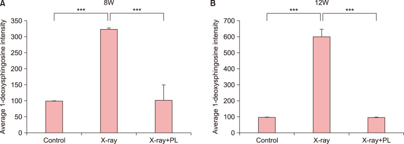
Fig. 8. Quantification of immunohistochemical analysis of 1-deoxysphinogosine in (A) 8-week-old (8W) and (B) 12-week-old (12W) hairless mice following X-ray and the physiological lipid mixture (PL) treatment. X-ray irradiated group showed significantly increased the accumulation of 1-deoxysphingosine in the skin compared to control in 8W and 12W hairless mice (p<0.001, p<0.001, respectively). The group treated with PL showed significantly lowered production of 1-deoxysphingosine in skin following X-ray irradiation compared to X-ray irradiation group without treatment of PL in 8W and 12W hairless mice (p<0.001, p<0.001, respectively). Expression levels of 1-deoxysphingosine staining were semiquantitatively analyzed using LASX software (Leica, Wetzlar, Germany). Results are expressed as mean±standard deviation of optical density of six different digital images. ***p<0.001.

Fig. 8