. 2019 Jul 1;31(4):403–413. doi: 10.5021/ad.2019.31.4.403

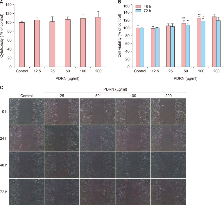
Fig. 1. Effects of polydeoxyribonucleotide (PDRN) on cytotoxicity and proliferation in human dermal fibroblast neonatal (HDF-n) cells. (A) The cytotoxicity of PDRN was measured using CCK-8 reaction solution in on HDF-n cells. The percentage of viable cells was expressed as a proportion of control cell viability, which was set to 100%. (B) The cell proliferation of HDF-n cells at 48 and 72 hours. The number of HDF-n cells treated with PDRN. (C) The cell migration of HDF-n. We observed these results with optical microscopes (CKX41; OLYMPUS, Tokyo, Japan) at each time point (0, 24, 48, and 72 hours). Significant differences from the control group are *p<0.05, **p<0.01.

Fig. 1