. 2019 Jul 1;31(4):454–457. doi: 10.5021/ad.2019.31.4.454

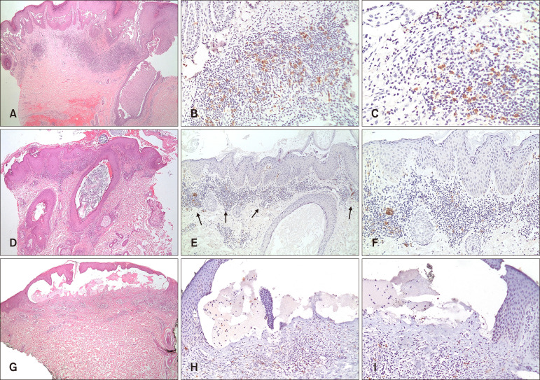
Fig. 1. (A~C) Pemphigus vulgaris. (A) Representative case showing suprabasal acantholysis with underlying dermal inflammatory infiltrate (H&E, ×40). (B, C) BDCA-2 immunostaining highlighted plasmacytoid dendritic cells (pDCs) in a superficial perivascular and interstitial distribution with pDCs making up more than 10% of the inflammatory infiltrate/pDC score of 2 (B: ×100, C: ×200). (D~F) Pemphigus foliaceus. (D) Representative case showing subcorneal acantholysis with underlying dermal inflammatory infiltrate (H&E, ×40). (E, F) BDCA-2 immunostaining highlighted pDCs in a superficial perivascular and interstitial distribution with pDCs making up more than 10% of the inflammatory infiltrate/pDC score of 2 (E: ×100, F: ×200). (G~I): Bullous pemphigoid. (G) Representative case with subepidermal blistering and underlying inflammatory infiltrate (H&E, ×100). (H, I) BDCA-2 immunostaining highlighted scattered pDCs in a perivascular and interstitial distribution with pDCs making up less than 10% of the inflammatory infiltrate/pDC score of 1 (H: ×100, I: ×100).

Fig. 1