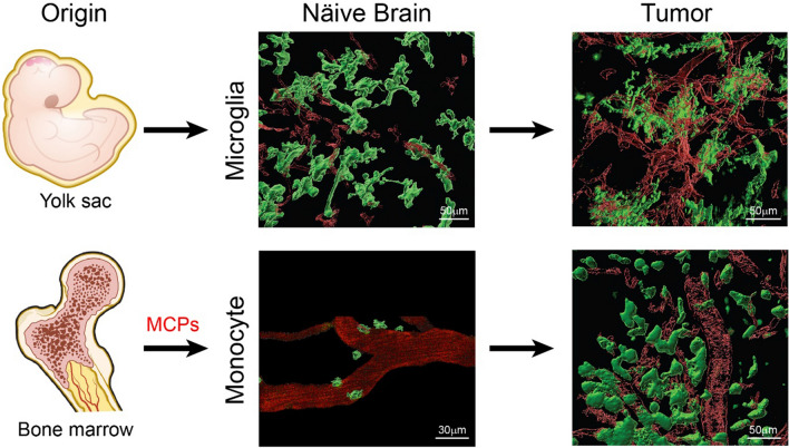
Fig. 1.
Myeloid cell lineage and morphology in healthy brain and glioblastoma. Microglia arise from yolk sac progenitors in the embryonic stage and reside permanently in the brain parenchyma. They appear as highly-ramified cells, but they rapidly change their morphology into an amoeboid-like shape in glioblastoma. Monocytes originate from the bone marrow and circulate in the blood, until they invade the brain when a glioblastoma emerges and differentiate into macrophages. They are round shaped while in circulation and upon infiltration into glioblastoma, making them morphologically indistinguishable from microglia. Images are taken from the murine glioblastoma model generated by using RCAS/tv-a, a somatic cell type-specific technology [41]. Reciprocal bone-marrow chimeras generated by using Cx3cr1-GFP mice allowed generation of only GFP-labeled bone marrow-derived macrophages or GFP-labeled microglia for imaging. Blood vessels are visualized with TRICT-dextran. Scale bars, 30 and 50 µm [41]

