Fig. 5.
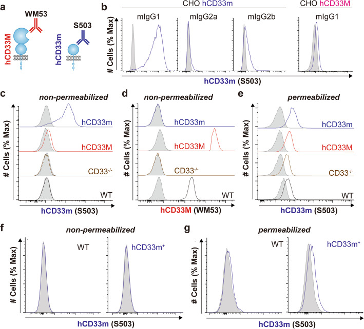
hCD33m drives a gain-of-function phenotype from an intracellular location. a Schematic demonstrating two anti-CD33 antibody clones. WM53 recognizes the N-terminal V-set domain unique to hCD33M while S503 recognizes a unique epitope on hCD33m. b S503 uniquely recognizes hCD33m-expressing CHO cells and is a mouse IgG1 antibody. Unstained S503 was incubated with cells CHO cells expressing hCD33m (left) or hCD33M (right), cell surface binding was detected with the indicated secondary antibody, and cells were analyzed by flow cytometry. c,d Cell surface expression of hCD33m and hCD33M with (c) S503 and (d) WM53, respectively, on WT or CD33−/− U937 cells transduced with EV (CD33−/−), hCD33M, or hCD33m. e Intracellular staining of hCD33m in U937 cells from WT or CD33−/− U937 cells transduced with EV (CD33−/−), hCD33M, or hCD33m. Cells were pre-blocked with unlabeled S503 followed by permeabilization of the cells prior to staining with AF647-labelled S503. f,g Expression of hCD33m from (f) non-permeabilized and (g) permeabilized primary microglia from WT and hCD33m+ mice

