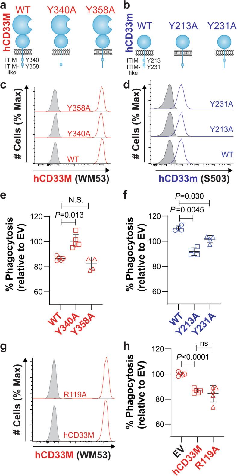
Fig. 6.

The gain-of-function role for hCD33m is signaling-dependent but not stem from loss of glycan-binding in hCD33M. a,b Cartoon diagram of the structure of (a) hCD33M and (b) hCD33m in the plasma membrane. The cytoplasmic tail of the proteins contains the ITIM and ITIM-like residues. Point mutants at each residue in each isoform were generated to test the importance of these residues. c,d Expression of (c) hCD33M (red) and (c) hCD33m (blue) and their associated mutants, transduced into CD33−/− U937 cells. For hCD33M (c), cell surface expression (non-permeabilized) was assessed. For hCD33m (d), intracellular expression (permeabilized) was assessed. Isotype-stained cells are shown in grey. e,f Phagocytosis of aggregated fluorescent Aβ1–42 in CD33−/− U937 (clone 4) cells virally-transduced to express (e) hCD33M or (f) hCD33m, along with their associated ITIM and ITIM-like mutants. % Phagocytosis represents the cytochalasin-D subtracted values referenced to the average of the empty vector (EV) cells set to 100%. (N = 4–5). g Cell surface expression of hCD33M and its R119A glycan-binding deficient mutant. Isotype-stained cells are shown in grey. h Phagocytosis of aggregated fluorescent Aβ1–42 in CD33−/− U937 cells (clone 4) transduced to express WT or R119A hCD33M. % Phagocytosis represents the cytochalasin-D subtracted values referenced to the average of the empty vector (EV) cells set to 100%. (N = 5)
