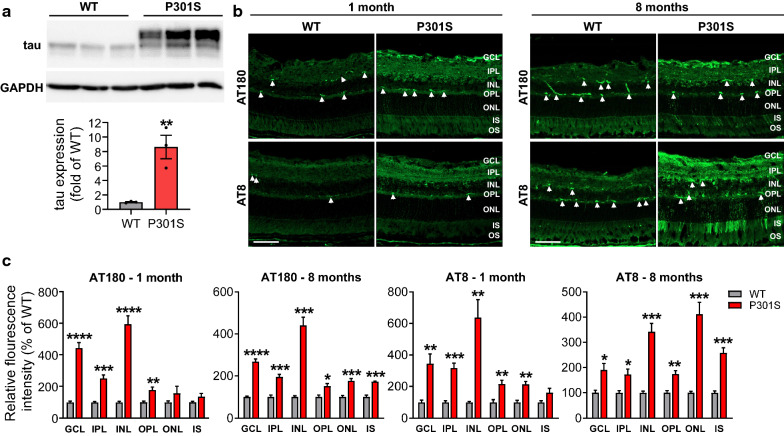
Fig. 1.
Total and phosphorylated tau are increased in the retina of P301S mice. a Retinal lysates from 3-month-old WT and P301S mice were blotted with anti-tau (Tau-5) for total tau. GAPDH was used as internal control. Graph represents densitometric analysis of tau protein normalized to GAPDH. n = 3/group. b Retinal sections from 1 and 8-month-old WT and P301S mice were stained with AT180 and AT8 antibodies for phosphorylated tau (green). Arrowheads indicate non-specific staining. c Quantification of fluorescence intensity of AT180 and AT8 in individual retinal layers. Non-specific staining was removed when performing quantification. Scale bar: 50 µm. n = 4/group. *p < 0.05; **p < 0.01; ***p < 0.001; ****p < 0.0001 versus WT. GCL ganglion cell layer, IPL inner plexiform layer, INL inner nuclear layer, OPL outer plexiform layer, ONL outer nuclear layer, IS inner segment, OS outer segment

