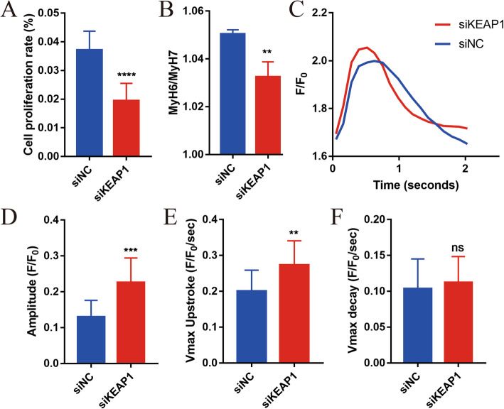
Fig. 5.
NRF2 promotes function maturation of hiPSC-CMs. a Quantification of the percentage of siKEAP1 and siNC hiPSC-CMs proliferating, as measured by EdU (each biological replicate n > 5, three biological replicates). b The proportion of MyH6 to MyH7 was decreased in the siKEAP1 hiPSC-CMs. n = 3. c, d HiPSC-CMs were analyzed by calcium transient kinetics with Fluo-4 AM (n > 3 cells per condition, three biological replicates): c representative calcium transient and d calcium transient amplitude (F/F0). e Maximum calcium transient upstroke velocities. f Maximum calcium transient decay velocities. The means ± SEM are shown. **P < 0.01, ***P < 0.001, ****P < 0.0001

