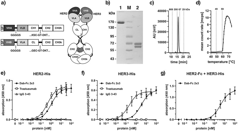
Figure 1.

A bivalent bispecific Dab-Fc antibody targeting HER2 and HER3. a) Schematic structure of a Dab-Fc fusion protein. b) SDS-PAGE of anti-HER2xHER3 Dab-Fc (Dab-Fc 2 × 3) under non-reducing (1) and reducing (2) conditions. Four micrograms of protein was loaded per lane, M = Marker. c) size-exclusion chromatography of purified Dab-Fc 2 × 3. d) determining melting points (Tm) by dynamic light scattering. e) binding of Dab-Fc 2 × 3 to immobilized HER2-His in ELISA. f) binding of Dab-Fc 2 × 3 to immobilized HER3-His in ELISA. Trastuzumab and IgG 3–43 were included as controls. g) Sandwich ELISA testing binding of HER3-His to titrated Dab-Fc 2 × 3 bound to immobilized HER2-Fc. Mean ± SD, n = 3
