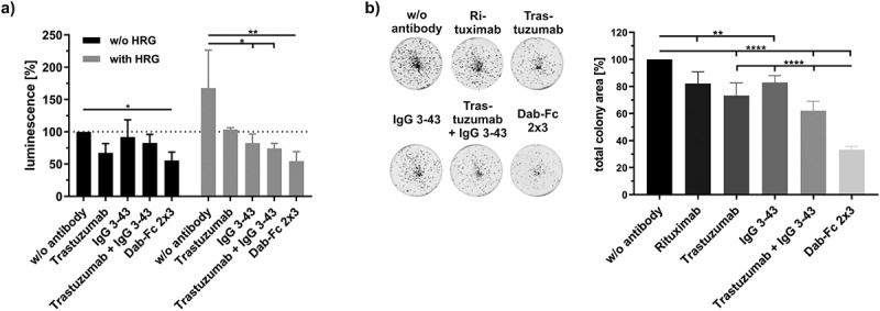
Figure 4.

Inhibition of proliferation and colony formation of NCI-N87 cells by Dab-Fc 2 × 3. a) Cells were seeded and on the next day starved with medium containing 0.2% FCS. After cultivating for 24 h, cells were incubated with the different antibodies (100 nM for Dab-Fc; 50 nM for the different antibodies) for 1 h and either left unstimulated (w/o HRG) or stimulated with HRG (25 ng/ml). After 7 days, cells were analyzed using cell titer glo 2.0. Data were normalized to the untreated and unstimulated cells. (n = 3; mean ± SD) statistics: one-way ANOVA. b) Cells were seeded in medium containing 10% FCS. After 24 h of cultivation, cells were treated with 50 nM of the different antibodies or with 100 nM with Dab-Fc molecule in medium containing 2% FCS. After 7 days, cells were retreated with fresh antibodies in 200 µl medium containing 2% FCS. After 12 days of incubation, cells were measured by crystal violet staining. Data were normalized to the untreated cells. Mean ± SD, n = 5, statistics: one-way ANOVA. *p < .05, **p < .01, ***p < .001, ****p < .0001
