. 2021 Jan 5;299(1):E204–E213. doi: 10.1148/radiol.2021203957

Figure 3g:

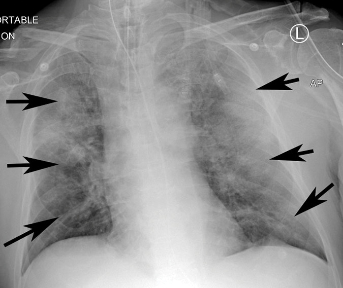
Example of CT scans and chest radiographs in the RSNA International COVID-19 Open Radiology Database. (a) Annotated axial CT image shows segmentation of characteristic bilateral multifocal ground-glass opacities in predominantly peripheral distribution (orange regions of interest). The CT image was classified as having typical appearance of coronavirus disease 2019 (COVID-19) pneumonia. (b) Annotated axial CT image shows segmentation of bilateral multifocal ground-glass opacities with diffuse distribution (orange regions of interest). The CT image was classified as having indeterminate appearance of COVID-19 pneumonia. (c) Thoracic CT image shows bilateral nodular and patchy opacities with peripheral and lower lung predominance involving four lung zones, annotated as typical for COVID-19 with moderate severity. (d) Thoracic CT image shows bilateral nodular and patchy opacities with peripheral and lower lung predominance involving more than four lung zones, annotated as typical appearance for COVID-19 and severe lung involvement. (e) Bedside chest radiograph with bilateral patchy and nodular opacities (arrows) with upper lung predominance involving more than four lung zones, annotated as indeterminate appearance for COVID-19 and severe lung involvement. (f) Bedside chest radiograph shows left lower lobe opacities (arrows) with small left pleural effusion involving a single lung zone, annotated as atypical appearance for COVID-19 and mild lung involvement. (g) Bedside chest radiograph shows bilateral patchy and nodular opacities (arrows) with upper lung predominance involving more than four lung zones, annotated as indeterminate appearance for COVID-19 and severe lung involvement. (h) Bedside chest radiograph shows left lower lobe opacity (arrow) with small left pleural effusion involving a single lung zone, annotated as atypical appearance for COVID-19 and mild lung involvement.

Example of CT scans and chest radiographs in the RSNA International COVID-19 Open Radiology Database. (a) Annotated axial CT image shows segmentation of characteristic bilateral multifocal ground-glass opacities in predominantly peripheral distribution (orange regions of interest). The CT image was classified as having typical appearance of coronavirus disease 2019 (COVID-19) pneumonia. (b) Annotated axial CT image shows segmentation of bilateral multifocal ground-glass opacities with diffuse distribution (orange regions of interest). The CT image was classified as having indeterminate appearance of COVID-19 pneumonia. (c) Thoracic CT image shows bilateral nodular and patchy opacities with peripheral and lower lung predominance involving four lung zones, annotated as typical for COVID-19 with moderate severity. (d) Thoracic CT image shows bilateral nodular and patchy opacities with peripheral and lower lung predominance involving more than four lung zones, annotated as typical appearance for COVID-19 and severe lung involvement. (e) Bedside chest radiograph with bilateral patchy and nodular opacities (arrows) with upper lung predominance involving more than four lung zones, annotated as indeterminate appearance for COVID-19 and severe lung involvement. (f) Bedside chest radiograph shows left lower lobe opacities (arrows) with small left pleural effusion involving a single lung zone, annotated as atypical appearance for COVID-19 and mild lung involvement. (g) Bedside chest radiograph shows bilateral patchy and nodular opacities (arrows) with upper lung predominance involving more than four lung zones, annotated as indeterminate appearance for COVID-19 and severe lung involvement. (h) Bedside chest radiograph shows left lower lobe opacity (arrow) with small left pleural effusion involving a single lung zone, annotated as atypical appearance for COVID-19 and mild lung involvement.