. 2021 Jan 5;299(1):E204–E213. doi: 10.1148/radiol.2021203957

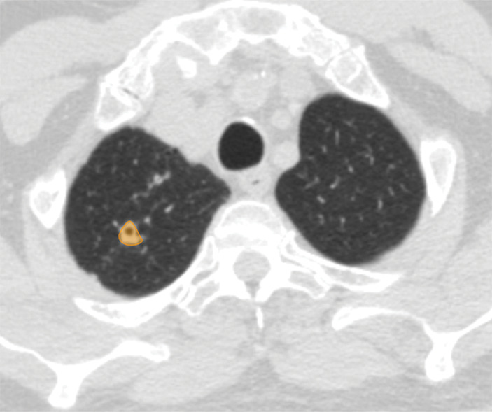
Figure 4c:

Examples of image-level annotations on axial CT images indicated with orange regions of interest. (a) Infectious opacity segmented in the left upper lobe. (b) Infectious tree-in-bud and/or micronodules segmented in the right lower lobe. (c) Infectious cavity segmented in the right upper lobe. (d) Noninfectious nodule or mass segmented in the posterior left pleura. (e) Atelectasis segmented in the left lower lobe. (f) Other noninfectious opacity segmented in the right lower lobe.

Examples of image-level annotations on axial CT images indicated with orange regions of interest. (a) Infectious opacity segmented in the left upper lobe. (b) Infectious tree-in-bud and/or micronodules segmented in the right lower lobe. (c) Infectious cavity segmented in the right upper lobe. (d) Noninfectious nodule or mass segmented in the posterior left pleura. (e) Atelectasis segmented in the left lower lobe. (f) Other noninfectious opacity segmented in the right lower lobe.