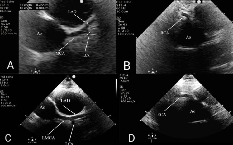
Figure 2.

Transthoracic echocardiography, parasternal short axis view. (A) Aorta (Ao) in cross-section with normal calibre LMCA, LAD and LCX. (B) Ao in cross-section and non-dilated, tapering RCA. (C) Dilated LMCA; dilated, non-tapering LAD and LCX. (D) Dilated, non-tapering RCA. LAD, left anterior descending coronary artery; LCX, left circumflex coronary artery; LMCA, left main coronary artery; RCA, right coronary artery.
