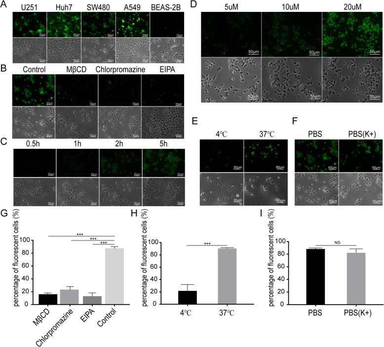
Fig. 2.
RGD4C penetration test. (a) The green fluorescence of EGFP was observed in SW480, U251, Huh7, and A549 tumor cells but not in normal BEAS-2B cells after incubation with RGD4C-EGFP for 5 h. (b)(g) When the endocytosis inhibitor MβCD, chlorpromazine or EIPA was added to a coculture of RGD4C-EGFP and SW480 cells, the numbers of fluorescent cells decreased significantly compared with the addition of the control reagent (P < 0.05). (c) SW480 cells were treated with 20 μM RGD4C-EGFP for 0.5 h, 1 h, 2 h or 5 h. Green fluorescence increased with increasing treatment time. (d) SW480 cells were treated at 37 °C for 5 h with 5 μM, 10 μM or 20 μM RGD4C-EGFP. The green fluorescence intensity of the SW480 cells increased gradually with increasing RGD4C-EGFP protein concentrations. (e) (h) The number of fluorescent cells was greater when incubated at 37 °C than when incubated at 4 °C. Incubation at 37 °C caused more RGD4C-EGFP to penetrate SW480 cells than incubation at 4 °C (p < 0.05). (f) (i) PBS and PBS rich in K+ were added to separate cocultures of SW480 cells and RGD4C-EGFP for 5 h. There was no significant difference in the percentage of fluorescent cells between the two groups

