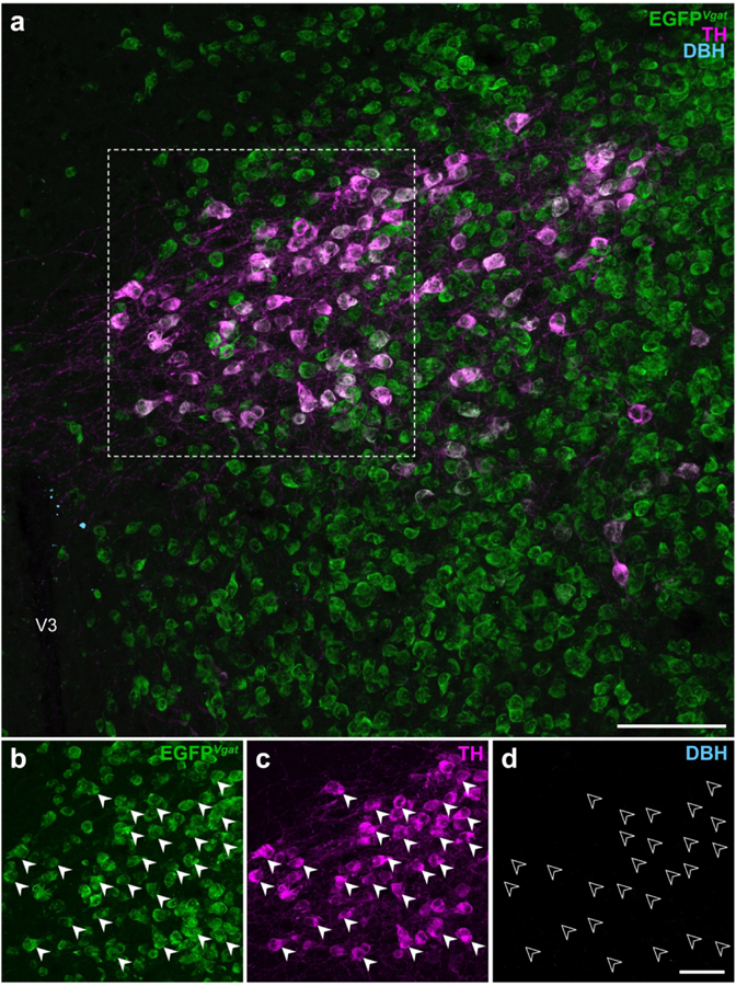
Figure 7. EGFPVgat TH-ir neurons in the ZI do not express dopamine β-hydroxylase.
Confocal photomicrographs from the ZI of Vgat-cre;L10-Egfp brain tissue (a) show EGFPVgat (b) in TH-ir neurons (c) but not immunoreactivity for dopamine β-hydroxylase (DBH; d). Filled arrowheads mark a representative sample of EGFPVgat TH-ir neurons (b, c) from the outlined area (a) that do not express DBH, as indicated by open arrowheads (d). Scale bars: 100 μm (a); 50 μm (b–d). V3, third ventricle.

