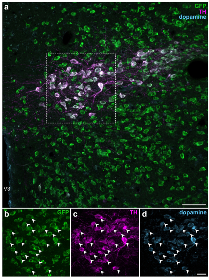
Figure 8. EGFPVgat TH-ir neurons in the ZI contain immunoreactivity for dopamine.
Confocal photomicrographs from the ZI of Vgat-cre;L10-Egfp brain tissue (a) show the colocalization of GFP (b), TH (c), and dopamine (d) immunoreactivities at high magnification within the same neuron. Filled arrowheads indicate representative GFP-, TH-, and dopamine-ir neurons. Scale bars: 50 μm (a); 20 μm (b–d). V3, third ventricle.

