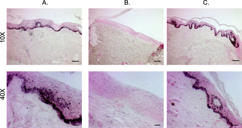
Fig 8. Fontana-Masson staining reveals melanin deposition of hyper- and hypo-pigmented patient HTS compared to normal skin.
Punch biopsies of distinct regions of hyper- (A), hypo- (B), and normally-pigmented (C) scar and skin were taken and were FFPE and Fontana-Masson stained. (Scale bar = 100 μm for 10X, top and 20 μm for 40X, bottom).

