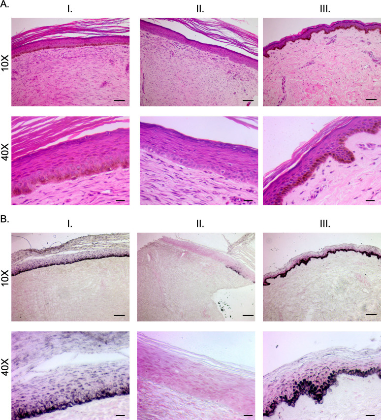
Fig 11. Staining reveals structural architecture and melanin deposition in hyper- and hypo-pigmented patient HTS compared to normal skin.
Punch biopsies of distinct regions of hyper- (I), hypo- (II), and normally-pigmented (III) scar and skin were taken and were FFPE and H&E stained (A). The same biopsies were Fontana-Masson stained (B). Images are from Subject #6 in Table 2. (Scale bar = 100 μm for 10X, and 20 μm for 40X).

