Figure 1.
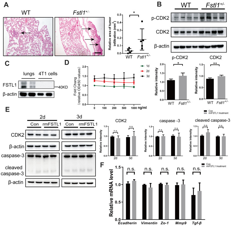
FSTL1 deficiency promoted growth of metastases in lung, while rmFSTL1 had no effect on 4T1 cells. The WT and Fstl1+/- female BALB/C mice were orthotropically transplanted with murine 4T1 cells. Fourteen days after inoculation, the mice were sacrificed for study. (A) H&E stained slices of lung from WT and Fstl1+/- tumor-bearing mice (n=5). Scale bar, 200 μm. Infiltrated tumor regions were measured by Image J software. (B) The protein levels of p-CDK2 and CDK2 in the lung tissues of WT and Fstl1+/- bearing-tumor mice (n=4). Densitometric measurement of band intensity normalized to that of β-actin. (C) The expression levels of FSTL1 protein in lung of WT mice and 4T1 cells. Densitometric measurement of band intensity normalized to that of β-actin. (D) 4T1 cells were treated with different concentrations of rmFSTL1 (0, 100, 200, 500, 1000 ng/mL) for 24h, 48h, or 72h and the cell viability was assessed by CCK-8 assay. (E) The expression levels of CDK2, Caspase-3 and cleaved Caspase-3 in 4T1 cells treated with 500 ng/mL rmFSTL1 for 48h or 72h. Densitometric measurement of band intensity normalized to that of β-actin. (F) The mRNA levels of genes related to EMT in rmFSTL1 treated groups and control groups, which was normalized to that of β-actin. Data are presented as mean ± SD. Each dot in the graphs represents an individual mouse. Data in the line chart represent three sets of independent experiments. n.s., not significant; *p < 0.05, **p < 0.01.

