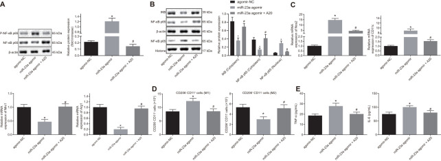
Figure 5.
Overexpression of miR-23a enhances inflammation response and M1 polarization of macrophages by inhibiting A20 and activating NF-κB. Macrophages were treated with exogenous miR-23a agomir (agomir-NC used as control) or exogenous miR-23a agomir and A20. (A) protein levels of p-NF-κB p65/NF-κB p65 in macrophages measured using Western blot analysis, normalized to β-actin; (B) protein levels of NF-κB p65 in the nucleus and cytoplasm measured using Western blot analysis, normalized to β-actin; (C) mRNA expression of Nos2, CD11c, Mrc1, and Arg1 determined using RT-qPCR in macrophages, normalized to β-actin; (D) M1 polarization of macrophages detected by flow cytometry; (E) levels of TNF-α and IL-6 measured by ELISA in macrophages. Values obtained from three independent experiments are expressed as mean ± SD and analyzed by one-way ANOVA followed by Bonferroni’s multiple comparison test among multiple groups. * p < 0.05 vs. macrophages treated with antagomir-NC plasmids; # p < 0.05 vs. macrophages treated with miR-23a agomir plasmids. Cell experiment was independently repeated for three times.

