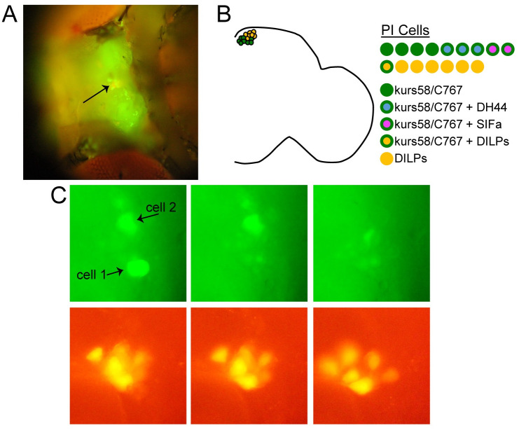
Fig 1. Single PI cell harvesting for RNA sequencing analysis.
(A) Fluorescence microscopy image of a UAS-nlsGFP /Dilp2mCherry; C767-GAL4/+ fly prepared for PI cell harvesting. Head cuticle between eyes has been removed, revealing the dorsal surface of the brain. Arrow points to PI region. (B) Schematic showing different PI cell types. On the left, one hemisphere of the fly brain is depicted. kurs58-GAL4 and C767-GAL4 (green circles) are largely restricted to the non-DILP-expressing PI neurons. DILP-expressing neurons are depicted in orange. The neurochemical makeup of the different PI cells is detailed on the right. In each hemisphere, there are ~7 DILP-expressing PI cells (orange cirlces) and ~10 kurs58/C767-GAL4-expressing PI cells (green circles). The neuropeptides DH44 and SIFa are present in non-overlapping populations of the kurs58/C767-GAL4-expressing cells. 3 of these cells express DH44 (green circles with blue interior); 2 express SIFa (green circles with magenta interior). (C) Closeup of the PI region showing two GFP-expressing PI cells (top) that were harvested for single-cell sequencing. Sequential images were taken before and after harvesting each cell. Nearby Dilp2mCherry-expressing cells were unaffected (bottom).

