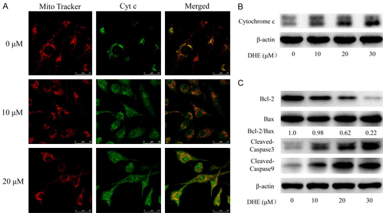
Figure 3.

DHE induced apoptosis through mitochondrial pathway in U87 cells. U87 cells treated with DHE at the indicated concentrations for 48 h. A. Laser scanning confocal microscope immunofluorescence analysis of cytochrome c (green) and mitochondria (red) co-localization in U87 cells. B. Cytoplasmic protein were extracted from the U87 cells and subjected to western blot analysis for cytochrome c and β-actin. C. And the levels of Bcl-2, BAX and cleaved caspase-3/9 proteins in total cell lysates from U87 cells were evaluated by western blot. The β-actin served as the protein loading control. Densitometric ratios of Bcl-2 and BAX proteins were quantified by Image J software.
