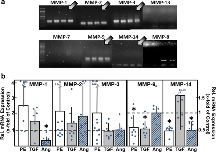
Fig. 2.
MMP mRNA expression in cardiomyocytes. (A) Total RNA was isolated from ventricular cardiomyocytes, RT-PCR performed, and amplification products were loaded on agarose gels (n = 4 independent culture preparations). mRNA expression of MMP types1, 2, 3, 9, and 14 was detected (indicated by arrows). (B) To determine the changes in mRNA expression under hypertrophic growth stimulation, cardiomyocytes were incubated for 24 h with phenylephrine (PE, 10 μM), TGFβ1 (1 ng/ml), or angiotensin II (Ang, 100 nM). mRNA expression of MMP types was analyzed by real-time RT-PCR (n = 7 – 9, *p < 0.05 vs. controls)

