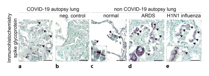
Fig. 3.
SARS-CoV‑2 detection methods in lung tissue. a Monoclonal anti-SARS spike glycoprotein antibody (mouse, monoclonal, Abcam, Cambridge, UK, Ab272420, 1:100) with apparent specific cytoplasmic granular staining pattern (a: arrowheads) in bronchial epithelia in autopsy lung tissue from a COVID-19-positive patient (SARS-CoV‑2 E [envelope protein] gene, RdRp [RNA-dependent RNA polymerase] gene, and N [nucleocapsid protein] gene positive in reverse transcriptase polymerase chain reaction [RT-PCR], disease duration 38 days). The regular negative control shows the lack of nonspecific binding of the secondary antibody (b: biotinylated horse anti-mouse, 1:300; scale bar = 40 µm). c–e Nonspecific binding of the primary antibody with similar granular staining pattern in bronchial epithelial cells and macrophages in autopsy lung tissue: c without any lung disease, d in acute respiratory distress syndrome (ARDS), and e in H1N1 influenza (scale bar = 40 µm)

