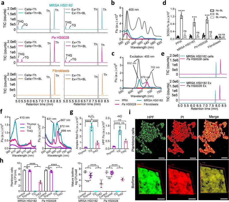
Fig. 6. Photo-oxidation of thymol to generate photosensitizers TQ and THQ exclusively in bacteria.
a UPLC-VION-IMS-QTOF-MS/MS analyses of thymol oxidation in cells (left) or extracts (right) of MRSA HS0182 (upper), Pa HS0028 (medium), or fibroblasts (bottom) treated with 0.5 mg/mL thymol in the absence or presence of 50 J/cm2 BL exposure. b and c Exciation (b) and emission (c) spectra of an indicated cellular extract in comparison with those of PPIX at 10 µM. d 1O2 is notably generated by 50 J/cm2 BL in MRSA HS0182, Pa HS0028, and PPIX solution but not in fibroblasts. 1O2 generation was blunted by NaN3 (BL + NaN3), a 1O2-specific quencher. e UPLC-VION-IMS-QTOF-MS/MS analyses of photo-oxidized thymol in the presence of NaN3 at 10 µM. BL illumination at 50 J/cm2, thymol at 0.5 mg/mL. f Exciation (left) and emission (right) spectra of thymol, TQ, and THQ each at 0.5 mg/mL. g H2O2 and •HO were generated by thymol, TQ, and THQ each at 0.2 mg/mL in combination with 50 J/cm2 BL. h Bactericidal activities of thymol, TQ, and THQ against planktonic cells (left) and established biofilms (right) of MRSA HS0182 and Pa HS0028 in the presence of 20 J/cm2 BL. The concentrations of three compounds were same at 0.05 mg/mL for planktonic cells and 0.1 mg/mL for established biofilms. i Representative fluorescence images of planktonic cells (upper) and established biofilms (bottom) of MRSA HS0182 after a lethal dose of the duo treatment. Bacterial viability and intracellular •HO were evaluated by PI and HPF staining, respectively. Scale bars: 2 µm in upper panel and 50 µm in bottom panel. All images in a, b, c, e, f, and i are representative of five independent experiments. Results in d, g, and h are presented as mean ± SD of at least five independent experiments. ****P < 0.0001; ***P < 0.001; **P < 0.01; *P < 0.05; and ns, no significance.

